
March 6, 2023
Zeptolab upgrade - March 2023
Thanks to the support of Zeptobars patrons - several vital hardware upgrades finally happened which will increase image quality and will make work more streamlined : custom wavelength RGB illuminator and 2-axis goniometer. Finally, we've also got Mastodon - @zeptobars@techhub.social - let's keep in touch there (just in case).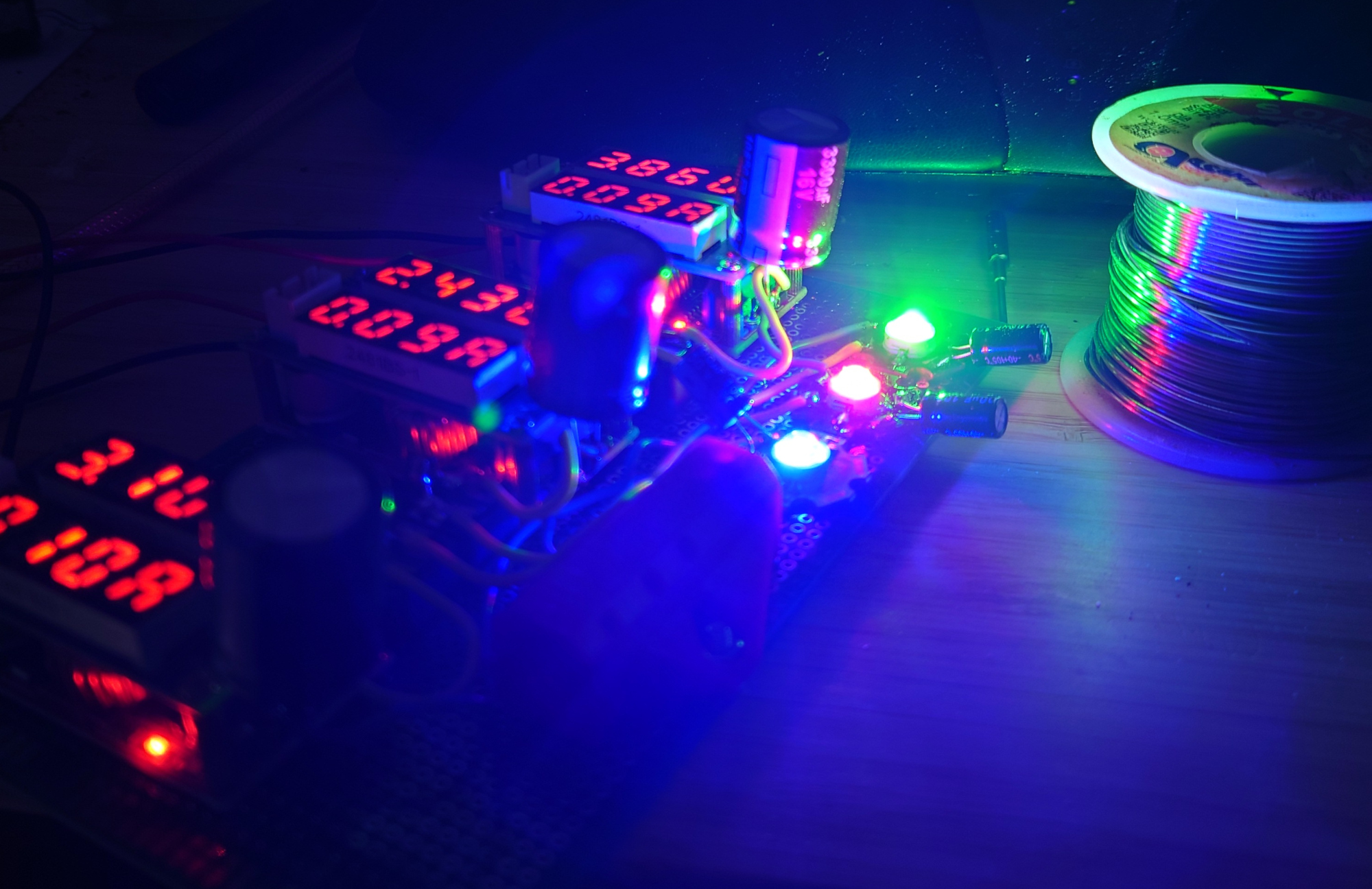
More details below:
First is custom RGB light source. I had to admit I've spent way too much on it due to fine-tuned wavelengths of the diodes.
My previous RGB light source that I used with old Chinese microscope used regular 450-520-620nm RGB leds. These do not match Bayer filter matrix in Sony imaging sensors very well (colors bleed into wrong channels reducing saturation and reducing efficiency of software correction of lateral chromatic aberration). So I decided to go for 440-545-660nm, which matches RGB filters and covers wider spectral range.
440nm is shortest wavelength that can be used on fluoride microscope lenses without significant quality degradation (they are designed for ~436nm and longer). Getting 440nm LED was straightforward - as it's just binning of commercially available GaN diodes. 5 batches from different suppliers was enough to nail down the wavelength.
660nm is the easiest. My very first batch was spot-on.
545nm diodes though are very challenging as they are in "green hole", where all LED semiconductors shows poorest quantum efficiency. In blue direction GaN starts working very well around 510-520nm. In yellow/red - multiple options starting at 580. I've tried to find extremely binned GaN diodes, and maybe 10 different batches gave me 530nm maximum. Phosphor-converted green LEDs have extremely wide spectrum, and definitely not suitable for this application. Finally, I've ordered GaP diodes from China, and in 4 batches one was perfect at 545nm. Surely, quantum efficiency is much lower, but I don't illuminate whole room anyways.
Per-channel adjustable current allows to set white balance to make sure gains of all RGB channels are 1.00 => lowest noise on the image. All extra capacitors (and inductors) - form CLC filter to ensure supply ripple does not introduce noise on progressive scan camera sensor.
LED light is side-coupled into 3mm optical fiber, which goes into microscope. I still want to build second version with the same LEDs with higher coupling efficiency. Otherwise I am already quite happy with it.
RGB illumination have 3 main benefits:
1) We work only on wavelength where lens shows highest quality,
2) RGB color channels are "independent", and correction of lateral chromatic aberration is much more efficient,
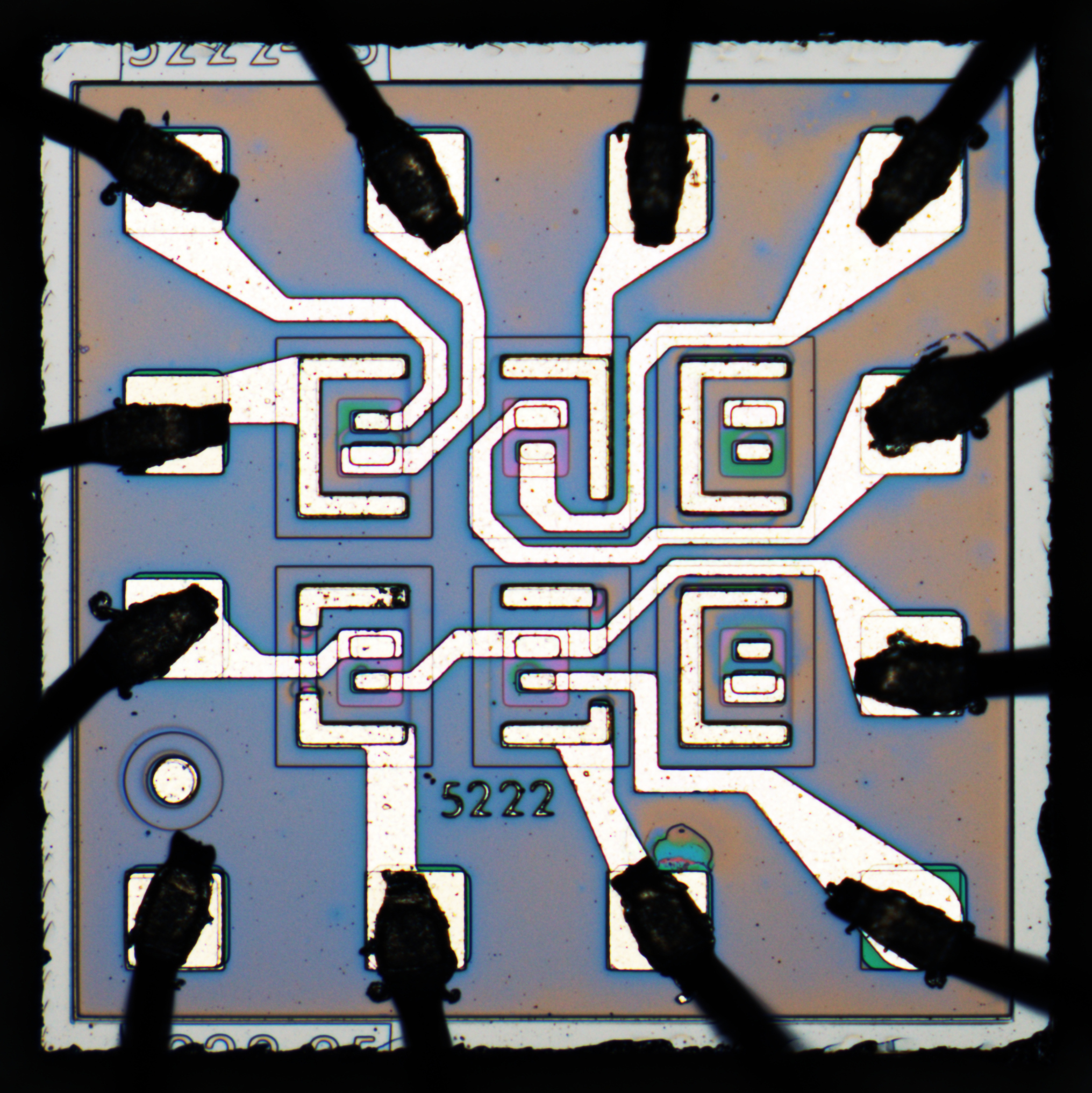
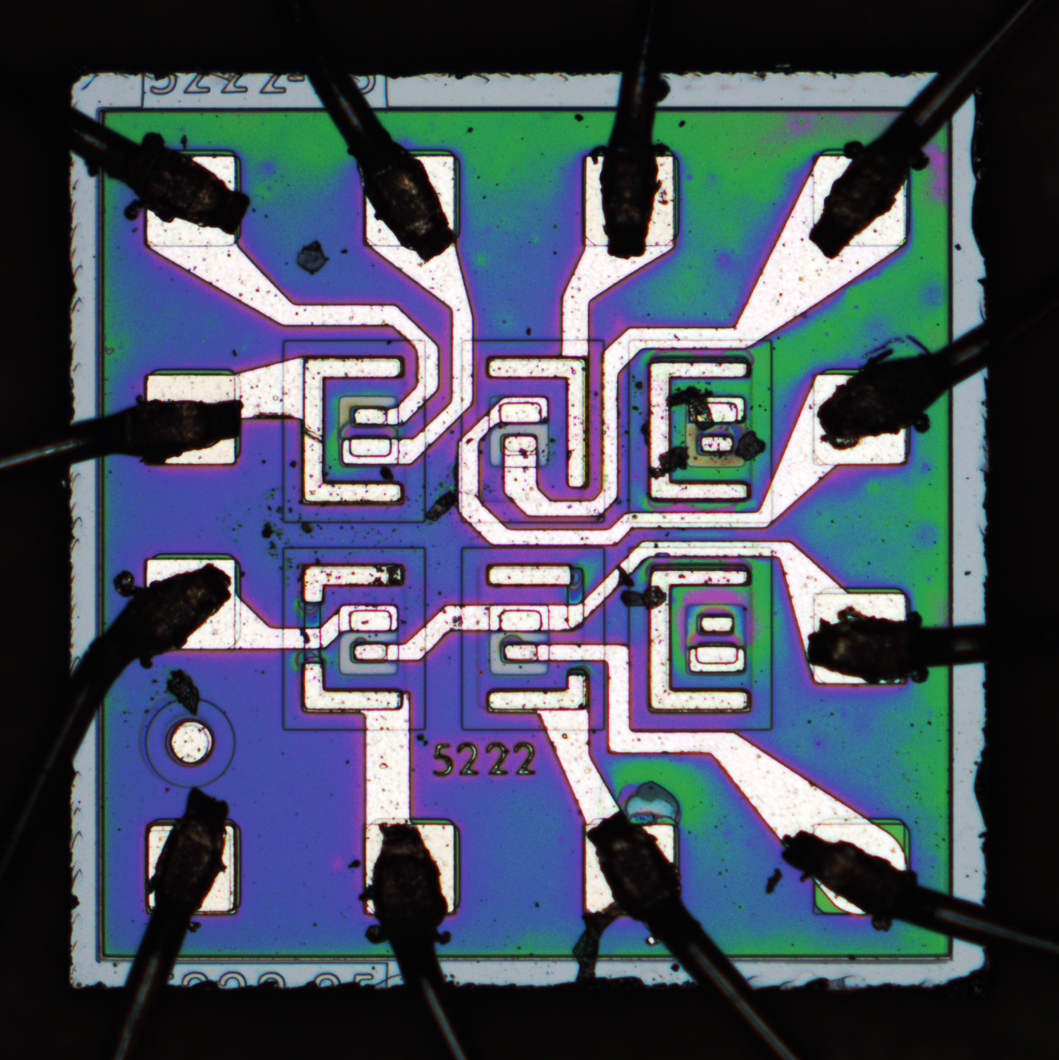
3) Color saturation is much higher without any special post-processing. See example below, exactly the same chip, similar post-processing but different light source:
White LED:
RGB LED:
2-axis goniometer:
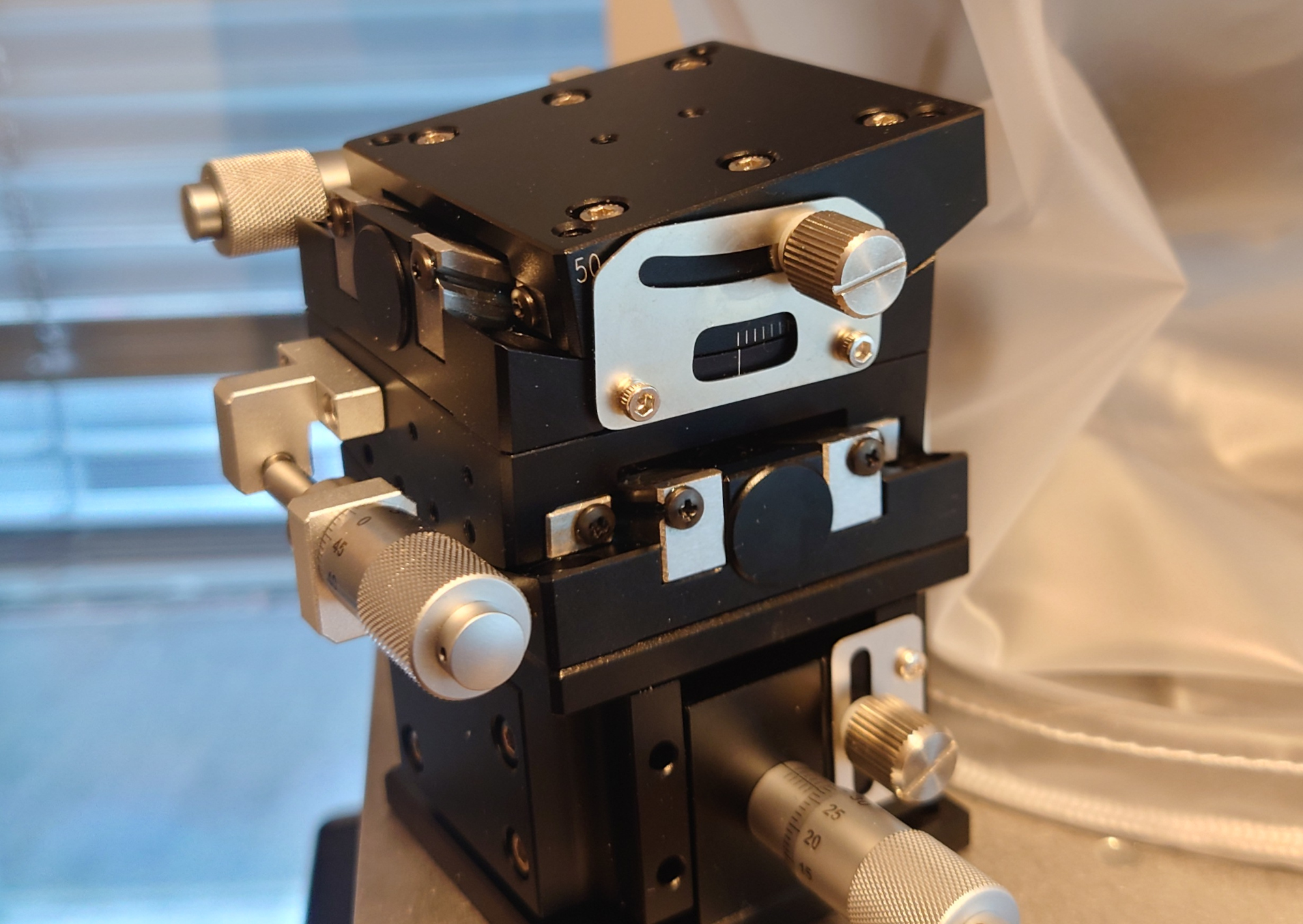
On the second upgrade - while ~80% of chips are flat enough to be photographed on regular XY stage, remaining ~20% are slightly tilted and became royal pain in the ass (typically in metal or metal-ceramic packages, due to uneven soldering or cases which impossible to lay flat). Dual-axis goniometer with micrometer adjustments from "ZY Automation" finally solves this issue.
You can also read previous article about how things worked in our lab in the past.


